Industrial CT Scanning Reveals Hidden Restoration Inside a Psittacosaurus Skull
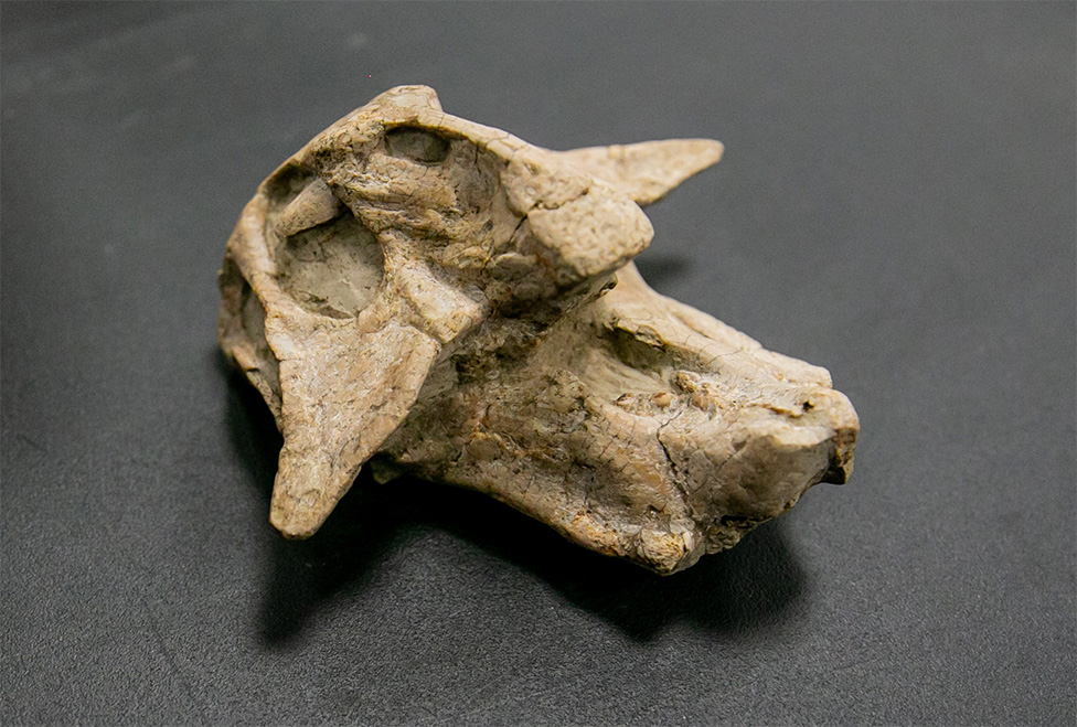
A collector submitted a Psittacosaurus skull fossil to Industrial Inspection & Consulting for non-destructive industrial CT scan evaluation. While the primary goal was to visualize the internal anatomy of the fossil, the scan quickly revealed something unexpected: historical restoration hidden beneath the surface. Without removing a single grain of material, the CT data differentiated authentic fossilized bone from modern repair materials, providing valuable insight into the specimen’s history and condition.
Fossils may undergo restoration before entering private collections or museums. External restoration can be nearly impossible to identify visually because repairs are intentionally blended into the surrounding fossil. Traditional inspection methods require destructive sampling or physical disassembly, neither of which is acceptable for valuable specimens. Industrial CT offers a completely non-destructive alternative.
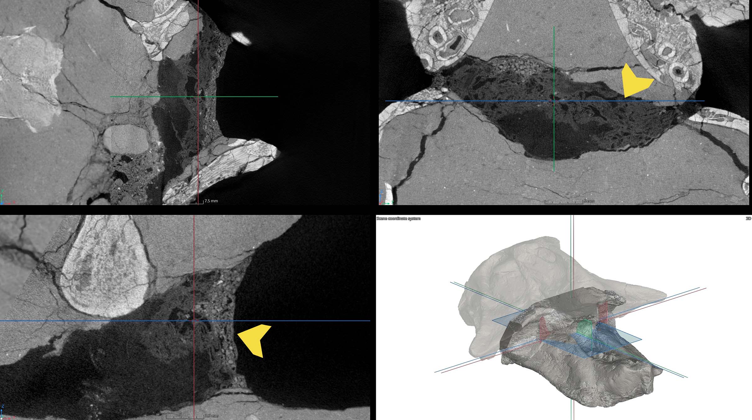
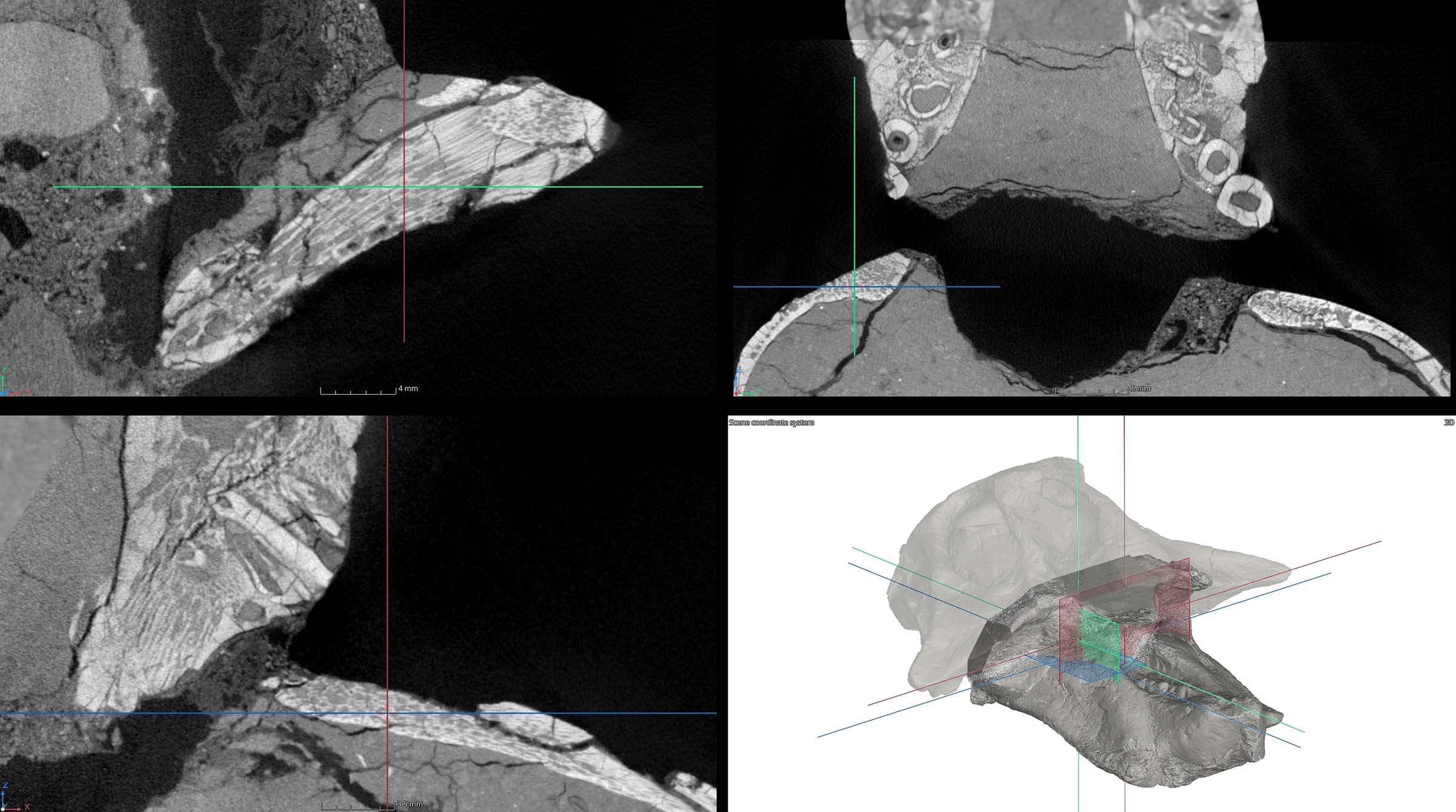
One of the most interesting discoveries was a repair material with a coarse, granular internal structure. Rather than presenting as fossilized bone, the material exhibited a dense, cement-like consistency throughout the repaired area.This strongly suggests that a mineral filler or restoration compound had been used during a previous conservation effort. The CT data showed a fibrous structure consistent with paper-based filler or paper mache used as an internal support beneath the reconstructed exterior. Although hidden from view, the material was immediately distinguishable from genuine fossil bone due to its dramatically different X-ray attenuation.
IIC's General Manager, Keith Irwin, discusses the psittacosaurus fossil project
Bone Segmentation from Surrounding Rock Matrix
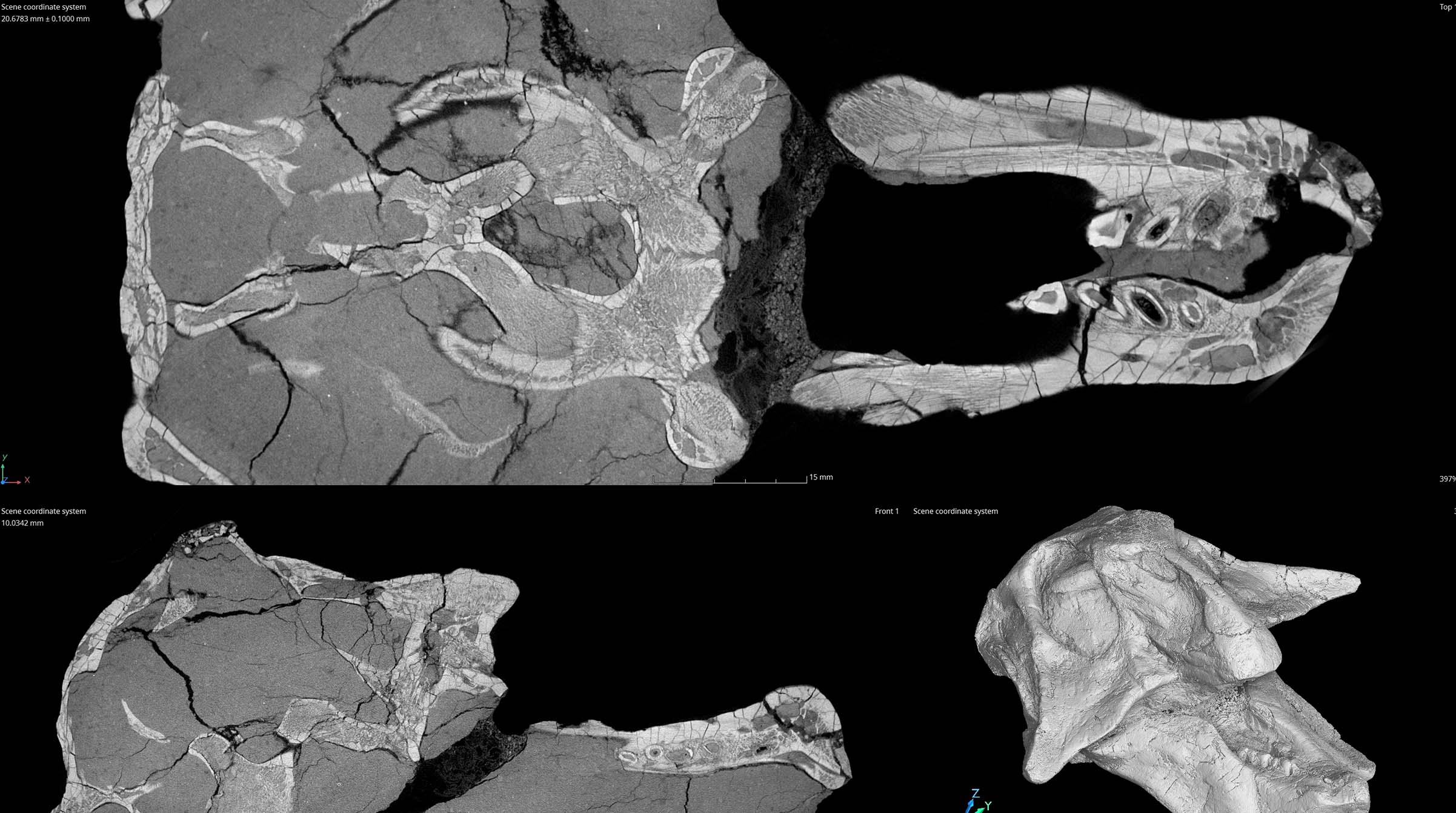
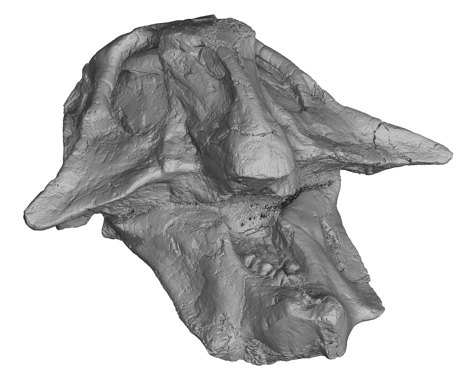
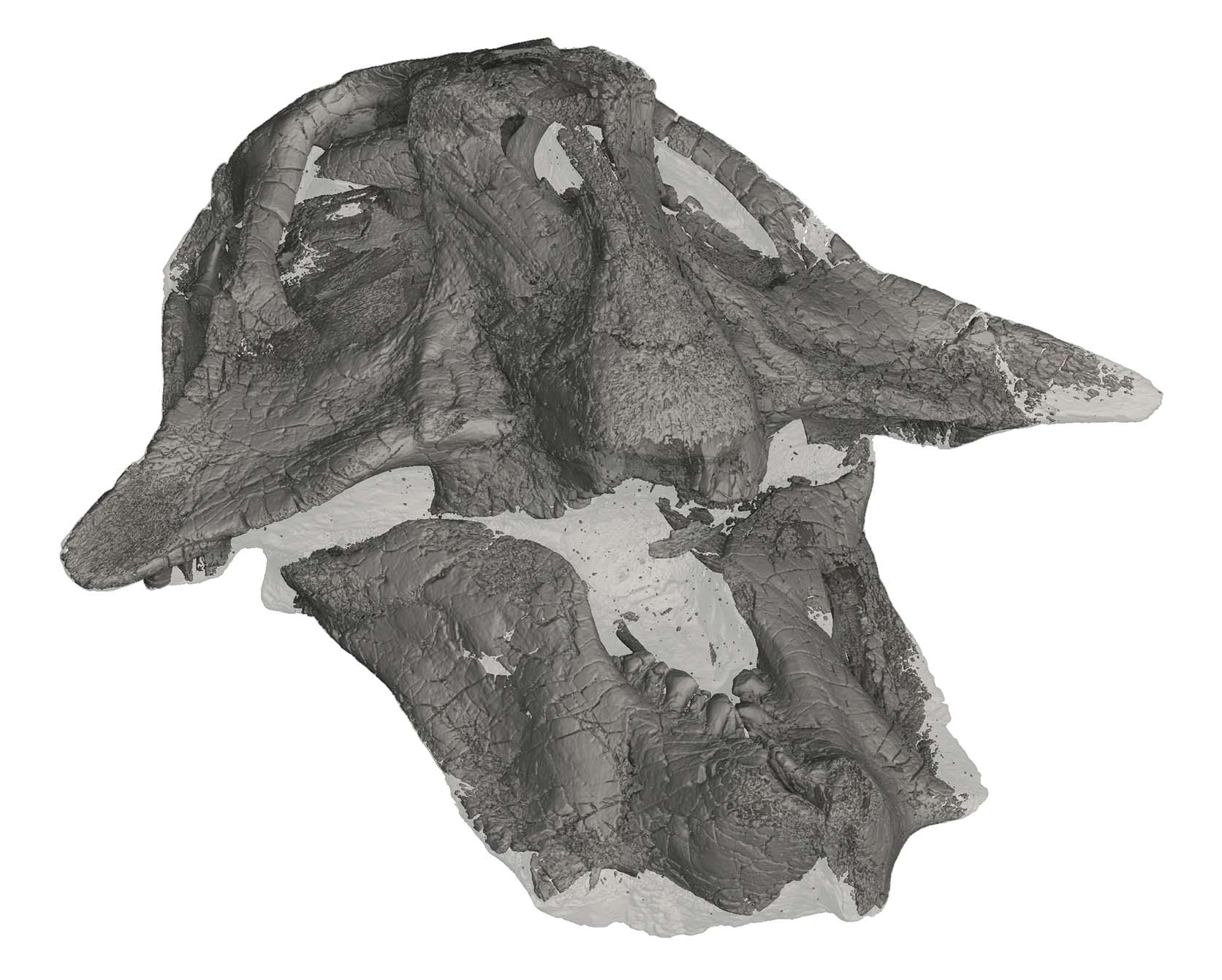
Using advanced volumetric analysis software, the fossilized bone was digitally segmented from the surrounding rock matrix. By separating materials based on their X-ray attenuation characteristics, we created a three-dimensional representation of the skull while isolating the remaining matrix.
Higher Resolution Imaging
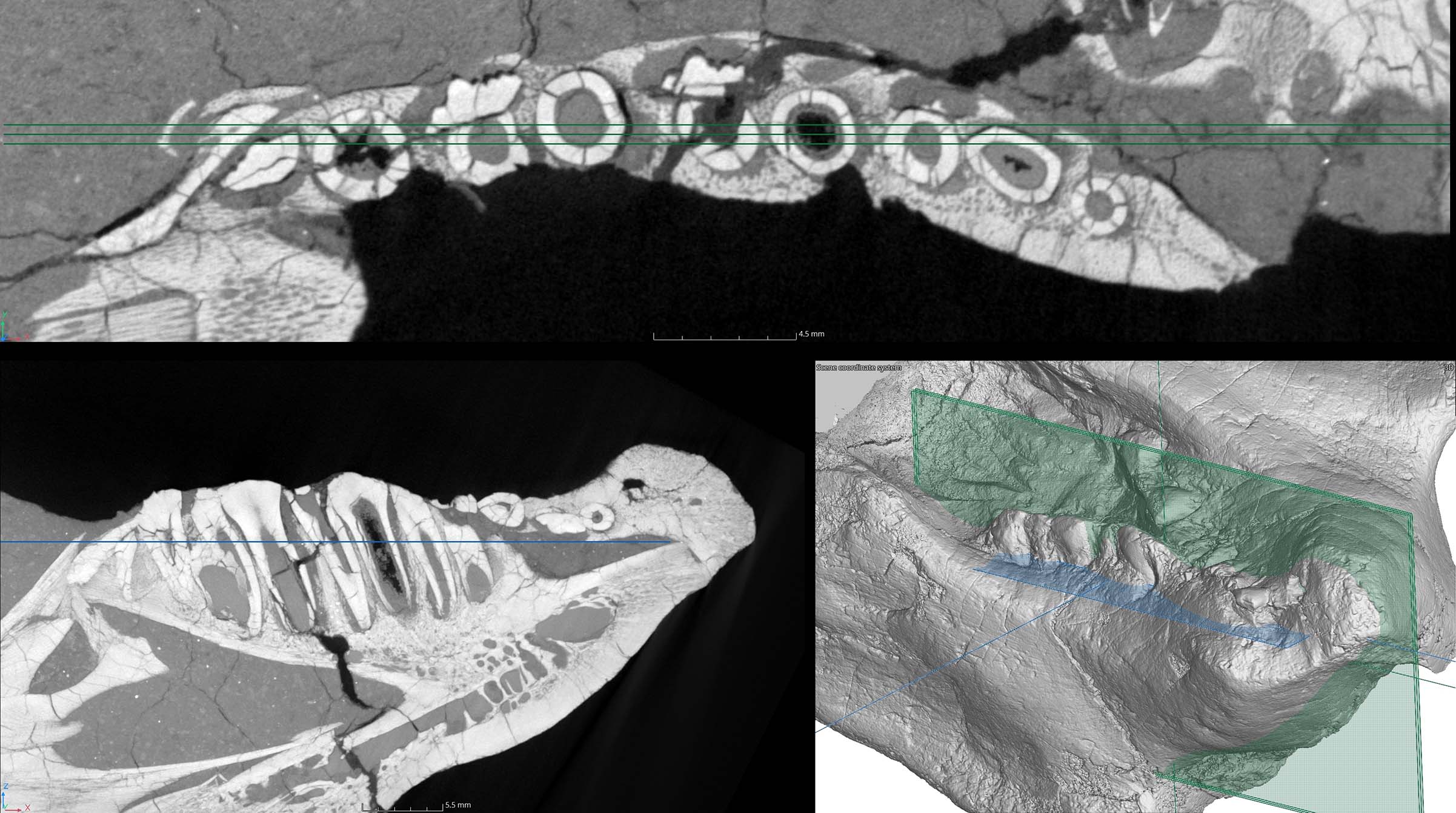
The scan didn’t just reveal hidden repairs. It exposed the fossil at a level impossible to achieve with visual inspection alone. A second scan was performed at 45um resolution, 2x higher than the initial scan, to capture individual bone striations, hairline fractures within the teeth, and the complete internal length of each tooth, including portions permanently hidden within the jaw. This allows researchers and collectors to study the specimen’s anatomy and condition in extraordinary detail without any destructive preparation.
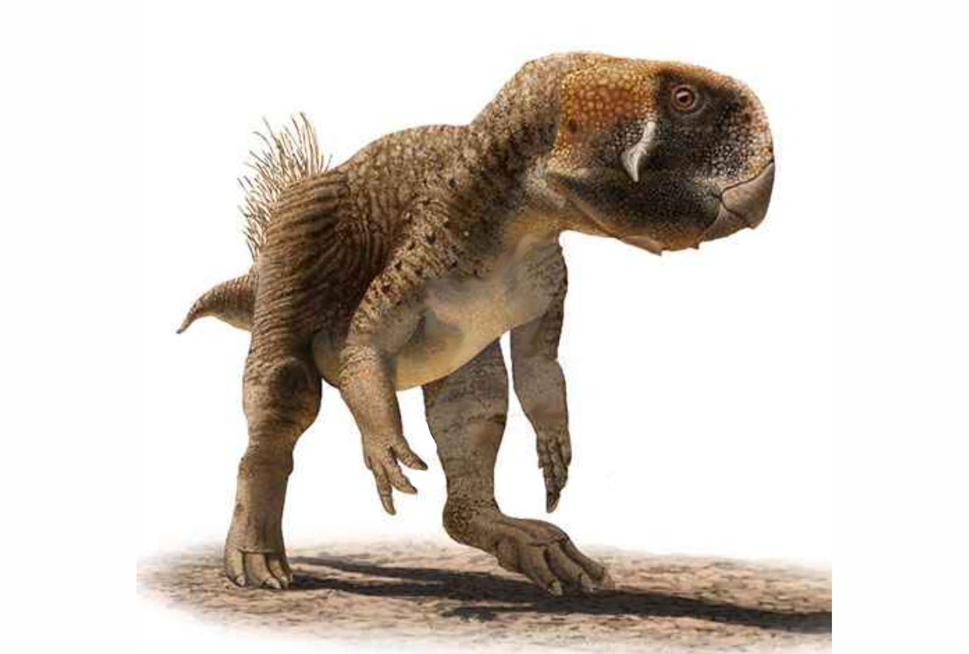
Psittacosaurs had self-sharpening teeth that were constantly replenishing through their lives, which would have been useful for cropping and slicing tough plant material. Unlike later ceratopsians, they did not have teeth suitable for grinding or chewing their food. Instead, they used gastroliths – stones swallowed to wear down food as it passed through the digestive system. Wikipedia Source