MICROFOCUS CT SCANNING OF FOSSILS
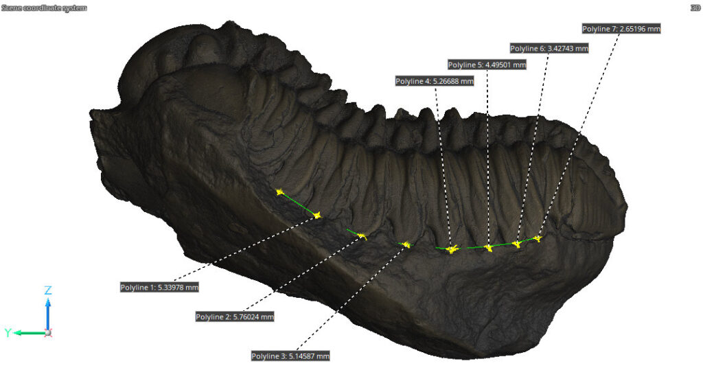
The oldest fossils are billions of years old. Traditional ways of examining fossils often employ destructive techniques which can compromise these timeless specimens. Industrial computed tomography (CT) scanning offers a non-destructive alternative that enables detailed examination of fossils’ internal and external structures. This case study explores how we can assist the field of paleontology with our microfocus CT scanning capabilities.
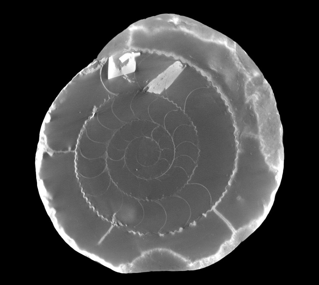
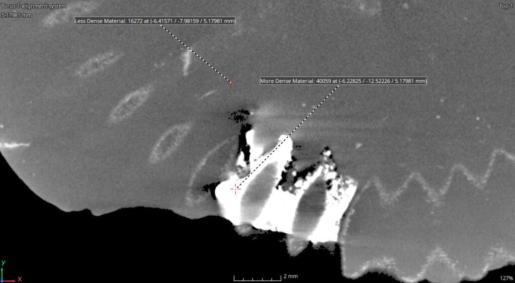
CT technology generates datasets by measuring the differential absorption of x-rays. Changes in material density and thickness cause differences in x-ray attenuation.
Depending on how a fossil was formed, there may be enough difference in material density for x-ray/CT to differentiate fossil from sedimentary rock.
The relative density is dependent on a variety of factors, including the types of minerals present in the fossil, the type of surrounding rock, and the conditions of fossilization. Each fossil and rock pairing can be unique, so the density comparison can vary.
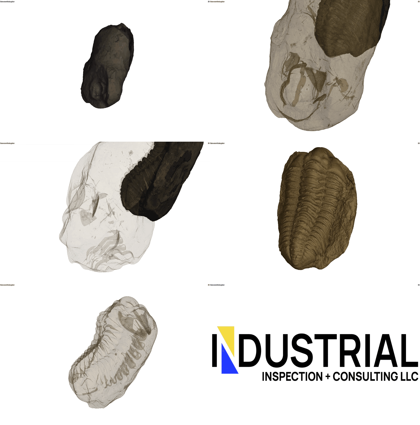
CT generates a 3D model by compiling thousands of 2D x-ray images. This means the 3D model has internal data and we are able to ‘slice’ through the test piece and look at it from any direction.
Using a high-end software for analysis and visualization of our microfocus CT data, we are able to perform a variety of measurements and analysis. We can generate videos for easy viewing and dissemination. We can create .stl models for 3D printing.
And in the case of these ammonites, the best part is we can do all of this analysis while the fossil remains undisturbed within the nodule.
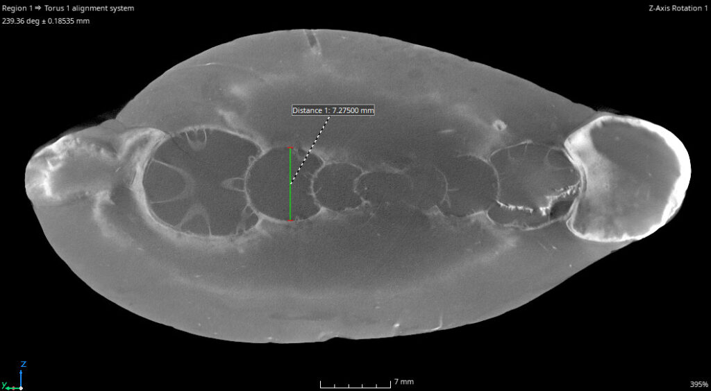
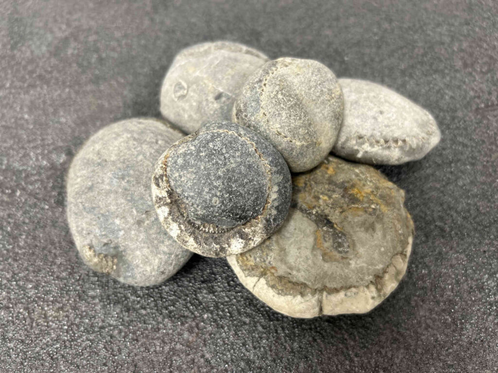
The application of industrial microfocus CT in fossil analysis allows for the detailed examination of internal structures without compromising the integrity of valuable specimens. By providing high-resolution, three-dimensional insights, micro-CT enables us to uncover previously inaccessible information about these ancient organisms. Contact us today to see how we can help with your fossils!
